AAEP 2020: Artificial Intelligence and Digital Radiography
Did you miss the live lecture on Artificial Intelligence in Veterinary Medicine? Don’t worry, we have it here for you to review!
Harnessing neural networks in veterinary medicine. See what its all about!
Explore why Vetel Diagnostics is the leading developer of artificial intelligence for veterinary diagnostic imaging by visiting our Intelligent Imaging page.
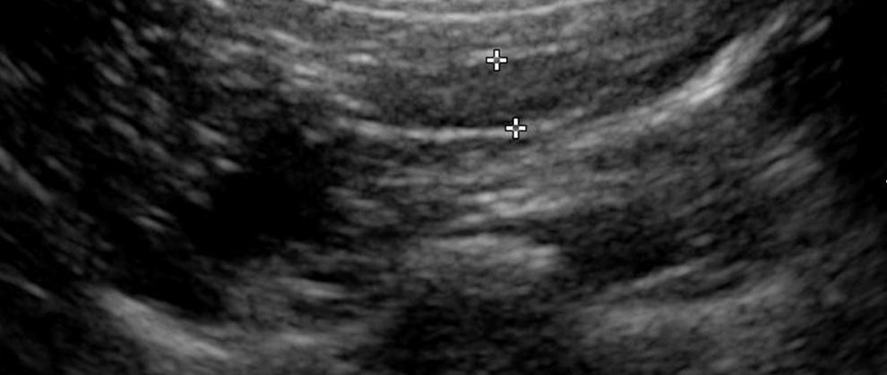
Imaging Essentials: Small Animal Abdominal Ultrasonography – A Tour of the Abdomen: Part 2
As originally published in Today’s Veterinary Practice,” March/April 2016 (Volume 6, Number 2)
Danielle Mauragis, AS, CVT, and Clifford R. Berry, DVM, Diplomate ACVRUniversity of Florida
Welcome to our series of articles on small animal abdominal ultrasonography. The first 3 articles provided an overview of basic ultrasonography principles, while the previous article and this one review a systematic pattern for scanning the abdomen. Future articles will discuss scanning principles for each abdominal organ and system, normal sonographic appearance of abdominal organs and systems, and identification of common abnormalities seen during
Never have to do a Vertebral Heart Score measurement by hand again
Vertebral Heart Score (VHS) for Companion Animals is now automated!
Our canine VHS has been independently verified by Dr. Sonya Gordon and the cardiology team at Texas A&M. In this study, the VHS measurements made by human experts for over 1000 dogs were compared with the measurement made by our AI, and the average difference was 3.3% which might be considered a statistical match.
See Vertebral Heart Score mark-up in action:
Our AI / Neural Network software platform is available in both a Cloud based application and also a version
embedded in our Metron software running on your local PC.
Find out more
Numbers Matter! Know what you’re looking for in a digital radiography system
When you’re in the market for a new digital radiography system, it can be hard to parse what metrics count most. Can you define the following?
DQE – Detector Quantum Efficiency
MTF – Modulation Transfer Function
lp/mm – line pairs per millimeter
If not, you’re in the majority! Watch this quick video to learn why these numbers are important.
Consider the numbers and take a look at some options that measure up.
More veterinary digital radiography solutions here.
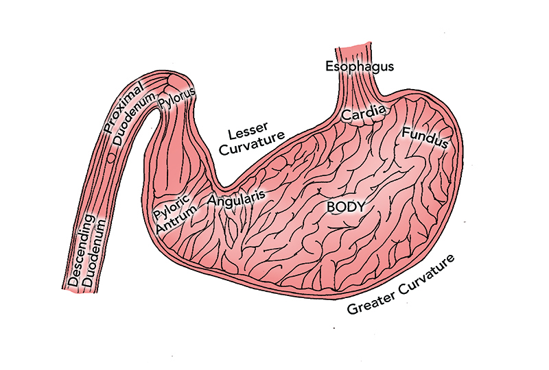
Endoscopy Essentials: Upper Gastrointestinal Endoscopy Techniques, Part 2
As originally published in Today’s Veterinary Practice,” March/April 2015 (Volume 5, Number 2)
Julie Callahan Clark, DVM, Diplomate ACVIM
University of Pennsylvania
Welcome to our newest column in Today’s Veterinary Practice—Endoscopy Essentials. Similar to our Imaging Essentials column, which addresses radiography by anatomic location, each article in this column will discuss endoscopic evaluation of a specifi c body system, reviewing indications, disease abnormalities, and proper endoscopic technique. The Endoscopy Essentials articles are archived at 
Upper gastrointestinal endoscopy (UGIE) is a minimally invasive procedure that can:
Aid in the diagnostic evaluation of clinical signs referable to
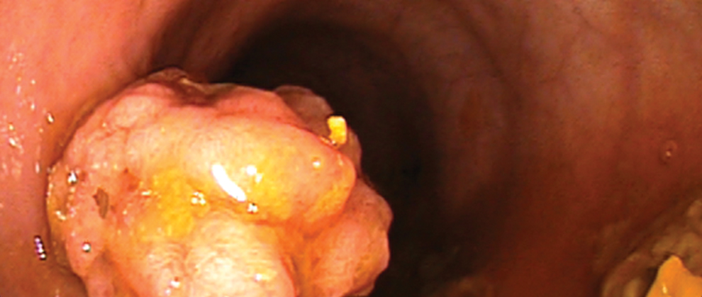
Endoscopy Essentials: Lower Gastrointestinal Endoscopy Series. Part 1: Overview of Lower Gastrointestinal Endoscopy
As originally published in Today’s Veterinary Practice,” July/August 2016 (Volume 6, Number 4)
Patrick S. Moyle, DVM, and Alex Gallagher, DVM, MS, Diplomate ACVIM University of Florida
Welcome to Endoscopy Essentials, a column that discusses endoscopic evaluation of specific body systems, reviewing indications, disease abnormalities, and proper endoscopic techniques. Visit tvpjournal.com to read the first three Endoscopy Essentials articles:
Overview of Upper Gastrointestinal Endoscopy (November/December 2014)
Upper Gastrointestinal Endoscopy Techniques (March/April 2015)
Endoscopic Foreign Body Retrieval (November/December 2015).
Lower gastrointestinal (GI) endoscopy is a minimally invasive diagnostic technique that allows the clinician to evaluate the mucosal surfaces of the rectum, colon,
Artificial Intelligence – Why should veterinarians care?
Artificial Intelligence (AI) can change the world of medicine. But how? And how will it affect the practice of veterinary medicine?
Deep learning is a type of AI in sophisticated software that utilizes neural networks, sometimes referred to as bioinformatics. When applied to diagnostic imaging, deep learning works by analyzing a bank of images to “learn” parts of the anatomy and judge what is considered “normal.” The more images the computer is given, the more precise its understanding becomes. Once the software learns what normal is, it can identify areas of
Press Release June 21, 2017: Vetel is Moving!
Vetel Diagnostics Outgrows Office of Eighteen Years
Veterinary Diagnostic Imaging Company Expands into New Location
San Luis Obispo, CA – Vetel Diagnostics, a leading veterinary diagnostic imaging company specializing in digital radiography systems, thermal imaging, ultrasound and endoscopes, has announced a move of its offices and manufacturing facilities to a new larger location just miles away from its original office of eighteen years.
Now located in the technology belt of San Luis Obispo, the office on Sacramento Drive has proven to be the perfect location to continue to grow
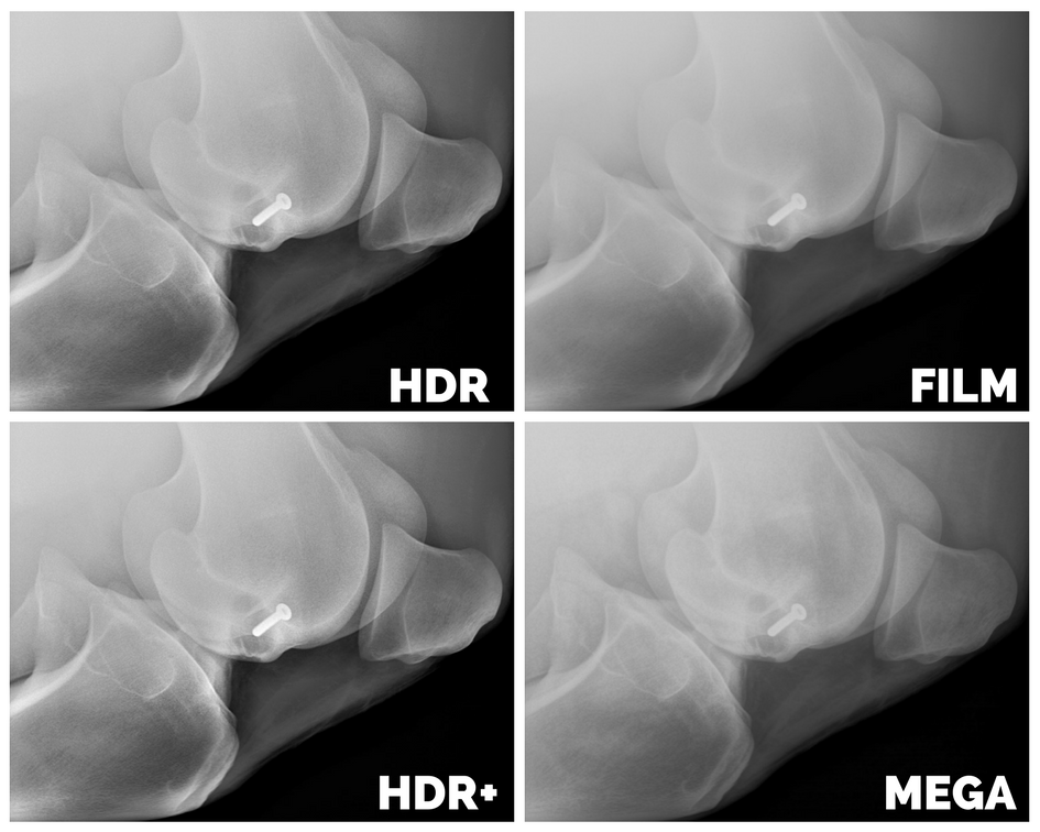
Anatomy of a Perfect Image: 3 Important Components of Image Quality
What affects image quality in flat panel detectors?
1. Pixel Pitch
Optimize the pixel pitch! (Remember, Pixel pitch describes the diameter of round pixels or the distance between dots <pixels> on a display screen. In digital radiography, the optimum pixel pitch is between 100-150 microns). A pixel pitch that is too small will give you blurred edge detail in the image anatomy, and also require a higher dose x-rays. A pixel pitch that is too high will produce a grainy image with poor spatial resolution or edge detail.
2. Scintillator
A scintillator is the part of the panel



Recent Comments